This Website and Domain Name are for sale.

Angiofibromas

What is Angiofibromas?

Angiofibromas are benign growths of blood vessels and fibrous tissue that can appear on the skin or mucous membranes. They can be associated with some genetic disorders or occur spontaneously. They are usually harmless and do not require treatment unless they cause cosmetic or functional problems.
dermatology-exam

What are the signs and symptoms of  Angiofibromas?

Some possible signs and symptoms of angiofibromas are:

  • Trouble breathing through one side of the nose or both sides due to airway obstruction
  • A lot of nosebleeds, usually with blood coming from only one nostril
  • A runny nose on one side that doesn’t go away after several days
  • Pain on the outer part of the elbow (lateral epicondyle) or with wrist movements
  • Vision, hearing, or speech issues due to tumor compression on the nerves or structures
  • Facial abnormalities such as droopy eyelids or bulging eyes
  • Loss of sense of smell (anosmia)

What are the causes of Angiofibromas?

The causes of angiofibromas are not well understood, but they may involve genetic mutations, hormonal factors, or local overgrowth of blood vessels and fibrous tissue. Some possible causes are:

  • Tuberous sclerosis: a genetic disorder that affects the skin, brain, kidneys, heart, and lungs. It is caused by mutations in the TSC1 or TSC2 genes, which encode proteins that regulate cell growth and division. Angiofibromas are one of the characteristic skin features of tuberous sclerosis, along with ash-leaf spots, shagreen patches, and ungual fibromas.
  • Birt-Hogg-Dubé syndrome: a rare genetic disorder that causes skin and kidney tumors, as well as lung cysts and pneumothorax. It is caused by mutations in the FLCN gene, which encodes a protein that interacts with other proteins involved in cell signaling and energy metabolism. Angiofibromas are one of the skin manifestations of Birt-Hogg-Dubé syndrome, along with fibrofolliculomas and trichodiscomas.
  • Multiple endocrine neoplasia type 1: a hereditary syndrome that leads to tumors in several endocrine organs, such as the parathyroid glands, the pituitary gland, and the pancreas. It is caused by mutations in the MEN1 gene, which encodes a protein that acts as a tumor suppressor. Angiofibromas are one of the cutaneous signs of multiple endocrine neoplasia type 1, along with collagenomas and lipomas.
  • Acquired angiofibroma: a benign growth that can occur spontaneously or due to unknown factors. It can appear on different areas of the body, such as the nose, face, penis, or mouth. It is composed of collagen, fibroblasts, and blood vessels. It is not associated with any genetic syndrome or systemic disease.

What treatments are available at the dermatologist for Angiofibromas?

Some treatments that are available at the dermatologist for angiofibromas are:

  • Laser therapy: This involves using a high-energy beam of light to destroy the blood vessels and fibrous tissue that make up the angiofibroma. It can reduce the size, color, and number of lesions, but may cause scarring, pain, or infection.
  • Abrasive therapy: This involves using a mechanical device to scrape off the surface layer of the skin where the angiofibroma is located. It can improve the appearance of the skin, but may cause bleeding, infection, or scarring. 
  • Topical mTOR inhibitors: This involves applying a cream or gel that contains a drug that blocks a protein called mTOR, which is involved in cell growth and division. It can shrink the angiofibroma and prevent new lesions from forming, but may cause skin irritation, infection, or allergic reactions. 
  • Topical beta-blockers: This involves applying a cream or gel that contains a drug that blocks the effects of adrenaline on the blood vessels. It can reduce the blood flow to the angiofibroma and make it less visible, but may cause skin irritation, infection, or allergic reactions. 
  • Surgical excision: This involves cutting out the angiofibroma with a scalpel or a sharp instrument. It can remove the lesion completely, but may cause scarring, pain, or infection. It may also require general anesthesia and stitches.
dermatology-consultation

Angiofibromas on the face

Angiofibromas on the face are also called fibrous papules. They are small, red to skin-colored bumps that usually appear on the nose or around the mouth. They are more common in middle-aged adults and can be mistaken for other skin conditions, such as basal cell carcinoma or molesSometimes, multiple facial angiofibromas can be a sign of a genetic syndrome, such as tuberous sclerosis, Birt-Hogg-Dubé syndrome, or multiple endocrine neoplasia type 1.

FAQ About Angiofibromas

How are angiofibromas diagnosed?

Angiofibromas are diagnosed by their clinical appearance and confirmed by histopathology. A skin biopsy may be performed to examine the tissue under a microscope and rule out other conditions that may look similar.

What are the complications of angiofibromas?

Angiofibromas may cause complications such as scarring, pain, infection, or recurrence after treatment. They may also be associated with systemic manifestations of genetic disorders, such as neurological, renal, endocrine, or pulmonary problems.

What is the prognosis of angiofibromas?

Angiofibromas have a benign course and do not transform into malignant tumors. However, they may persist or recur after treatment. The prognosis also depends on the presence and severity of systemic involvement in genetic disorders.

Are angiofibromas contagious?

No, angiofibromas are not contagious. They are not caused by an infection or transmitted by contact.

Is there a dermatologist near me in Austin that offers treatment for angiofibromas?

Yes. At our Austin dermatology office we offer treatment for angiofibromas to patients from Austin and the surrounding area. Contact our office today to schedule an appointment.

Lichen Planus

What is Lichen Planus?

Lichen planus is a condition that affects the skin, hair, nails, mouth and genitals. It causes purple, itchy, flat bumps on the skin and white patches or sores in the mouth and genitals. It is an autoimmune disorder, which means the immune system attacks the cells of the skin and mucous membranes. The exact cause of lichen planus is unknown, but it may be related to genetic factors, stress, infections, allergies or drugs. Lichen planus is not contagious and cannot be passed from one person to another. It can be treated with medicines and other therapies to relieve the symptoms and prevent complications.

dermatology-exam

What are the signs and symptoms of Lichen Planus?

 Some of the common signs and symptoms of lichen planus are:

  • Purple, shiny, flat bumps on the skin, often on the inner forearms, wrists or ankles. These bumps may have tiny white lines running through them, called Wickham’s striae.
  • Itching, pain or blisters on the affected skin areas.
  • Lacy white patches, redness, swelling, peeling or sores on the mouth and genitals.
  • Ridges, grooves, splitting, thinning or loss of nails.
  • Redness, irritation, tiny bumps, thinning hair, patches of hair loss or scars on the scalp.

What are the causes of Lichen Planus?

The causes of lichen planus are not fully understood, but it is believed to be related to the immune system. Lichen planus is a condition where the immune system mistakenly attacks the cells of the skin or mucous membranes, causing inflammation and rash. The reason why the immune system does this is unknown, but it may be influenced by genetic factors, environmental factors, or certain triggers. Some of the possible triggers of lichen planus are:

  • Hepatitis C infection.
  • Pain relievers and other medicines.
  • An allergic reaction to the metal in dental fillings.

Lichen planus is not contagious and cannot be passed from one person to another.

What treatments are available at the dermatologist for Lichen Planus?

Some of the treatments that are available at the dermatologist for lichen planus are:

  • Corticosteroids: These are medicines that reduce inflammation and redness. They can be applied to the skin as creams or ointments, or taken as pills or injections.
  • Oral anti-infection drugs: These are medicines that fight infections that may trigger or worsen lichen planus. Examples are hydroxychloroquine (an antimalarial drug) and metronidazole (an antibiotic).
  • Antihistamines: These are medicines that relieve itching by blocking the effects of histamine, a chemical released by the immune system during an allergic reaction.
  • Light therapy: This is a treatment that exposes the affected skin to ultraviolet B light, which can help clear up lichen planus. It may cause some side effects such as skin color changes or sunburn.
  • Tacrolimus ointment or pimecrolimus cream: These are medicines that are used to treat eczema, another skin condition. They may also help with lichen planus by suppressing the immune system and reducing inflammation.
dermatology-consultation

FAQ About Lichen Planus

How is lichen planus diagnosed by a doctor or a dermatologist?

Lichen planus is diagnosed by a doctor or a dermatologist based on the appearance of the skin or mucous membranes and the medical history of the patient. The doctor may also perform some tests to confirm the diagnosis or rule out other conditions that may look similar to lichen planus.

Who is at risk of getting lichen planus?

Anyone can develop lichen planus, but it is more common in middle-aged adults, especially women. It is also more likely to affect people who have a family history of the condition, or who have other autoimmune diseases.

Is lichen planus contagious?

 No, lichen planus is not contagious. You cannot catch it from or spread it to another person.

Is lichen planus related to cancer?

Lichen planus is not a cancerous condition, but it may increase the risk of oral or genital cancer in rare cases. Therefore, it is important to have regular check-ups with your doctor if you have lichen planus of the mouth or genitals.

Is there a dermatologist near me in Austin that offers treatment for Lichen Planus?

Yes. At our Austin dermatology office we offer treatment for Lichen Planus to patients from Austin and the surrounding area. Contact our office today to schedule an appointment.
Skin Tags

What are Skin Tags?

Skin tags are small pieces of soft, hanging skin that may have a stalk. They are benign, noncancerous, growths that can appear anywhere on your skin, but often develop on the neck, eyelids, or underarms. They are usually the same color as your skin or a little darker. They are not dangerous, but they can be removed for aesthetic or medical reasons.

dermatology-exam

What do Skin Tags look like?

The signs and symptoms of skin tags are:

  • Small pieces of skin that may be the same color as surrounding skin or darker
  • Soft and hanging growths that may have a stalk
  • Usually painless and harmless, unless they get irritated or bleed
  • Commonly found in areas where skin rubs against skin, such as eyelids, armpits, neck, groin, or underarms

What treatments are available at the dermatologist for Skin Tags?

There are two main methods that dermatologists use to remove skin tags:

  • Cryosurgery: This involves freezing the skin tag off with liquid nitrogen, which is very cold and destroys the skin tag. This works well for small skin tags, but may cause some discomfort, blistering, or scarring.
  • Snipping: This involves cutting the skin tag off with a sterile scissors or scalpel, usually after numbing the area with a local anesthetic. This works well for larger or multiple skin tags, but may cause some bleeding, infection, or scarring.
older-man

Skin Tags vs Moles

  • Skin tags are soft and hanging growths that usually have a stalk that connects them to the skin. They are usually flesh-colored or a little darker, and can develop anywhere on the skin, especially in areas where there is friction. They are benign and harmless, but may be removed for cosmetic or medical reasons.
  • Moles are flat or slightly raised growths that are usually round and one color. They can be brown, pink, red, black, blue, tan, or skin tone. They can develop anywhere on the skin, but are more common on sun-exposed areas. They are usually benign, but some may be precancerous or cancerous, and should be checked by a dermatologist regularly.

FAQ About Skin Tags

Are skin tags dangerous or contagious?

No, skin tags are benign (noncancerous) and do not pose any health risks. They are not contagious and cannot spread from one person to another.

How are skin tags diagnosed?

Skin tags are usually diagnosed by their appearance and location on the skin. However, some skin conditions or cancers can mimic the look of skin tags, so it is important to have them checked by a dermatologist to rule out any serious problems.

Can skin tags turn into cancer?

No, skin tags are benign and do not turn into cancer. However, some skin cancers can look like skin tags, so it is important to have any suspicious growths checked by a dermatologist to rule out any serious problems.

Is there a dermatologist near me in Austin that offers treatment for Skin Tags?

Yes. At our Austin dermatology office we offer treatment for Skin Tags to patients from Austin and the surrounding area. Contact our office today to schedule an appointment.
Mosquito Bites

Mosquito Bites

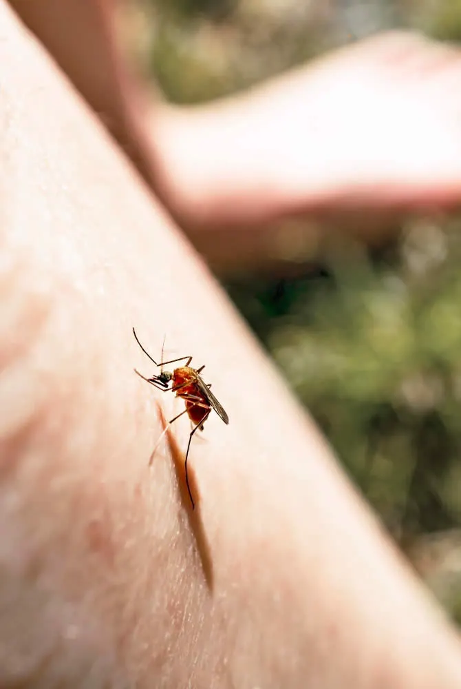
Mosquito bites are the itchy bumps that form on the skin after mosquitoes feed on your blood. Mosquitoes are insects that have a special mouthpart (proboscis) that can pierce the skin and suck blood. As they do so, they inject saliva into the skin, which causes an immune system reaction. This reaction results in swelling, redness and itching. Some people may have more severe reactions to mosquito bites, such as fever, hives or infection.

dermatology-exam

What are the signs and symptoms of Mosquito Bites?

Some of the signs and symptoms of mosquito bites are:

  • A puffy and reddish bump that appears a few minutes after the bite
  • A hard, itchy, reddish-brown bump, or multiple bumps that appear a day or so after the bite or bites
  • Small blisters instead of hard bumps
  • Dark spots that look like bruises
  • A large area of swelling and redness
  • Low-grade fever
  • Hives
  • Swollen lymph nodes

These symptoms may vary depending on the person’s immune system reaction and the type of mosquito that bites them. Some people may have more severe reactions than others, especially children and people with immune system disorders. 

What treatments are available at the dermatologist for Mosquito Bites?

Some of the treatments that are available at the dermatologist for mosquito bites are:

  • Topical corticosteroids: These are creams or ointments that contain steroids that can reduce inflammation and itching. They are usually prescribed for severe or persistent reactions, or for people who have skin conditions like eczema or psoriasis that can worsen with mosquito bites. 
  • Oral antihistamines: These are pills that block the effects of histamines, which are chemicals that cause itching and swelling. They can help relieve the symptoms of mosquito bites, especially if taken before exposure to mosquitoes. 
  • Oral corticosteroids: These are pills that contain steroids that can suppress the immune system and reduce inflammation. They are usually prescribed for very severe or widespread reactions, or for people who have a history of anaphylaxis (a life-threatening allergic reaction) to mosquito bites. 
  • Allergy shots: These are injections that contain small amounts of mosquito saliva or other allergens that can trigger an immune response. They are given over a period of time to gradually desensitize the body to the allergens and reduce the severity of future reactions.
mosquito-sits-on-arm

FAQ About Mosquito Bites

What are the complications of mosquito bites?

Mosquito bites can cause complications such as infection, scarring, and disease transmission.

How long do mosquito bites last?

The duration of mosquito bites depends on several factors, such as the person’s immune system reaction, the type of mosquito that bites them, and the treatments they use. 

Is there a dermatologist near me in Austin that offers treatment for Mosquito Bites?

Yes. At our Austin dermatology office we offer treatment for Mosquito Bites to patients from Austin and the surrounding area. Contact our office today to schedule an appointment.

Milia

What is Milia?

Milia are small, white or yellowish bumps that appear on the skin, usually on the face. They are caused by keratin, a protein that normally protects the skin, getting trapped under the skin and forming tiny cysts. Milia are not harmful or contagious, but they can be annoying or cosmetically undesirable for some people.

dermatology-exam

What are the signs and symptoms of Milia?

The signs and symptoms of milia are:

  • Small, white or yellowish bumps on the skin, usually on the face.
  • Bumps are dome-shaped, hard, and measure 1 to 2 millimeters in diameter.
  • Bumps are not painful or itchy, and do not cause inflammation or infection.
  • Bumps can appear in groups or clusters, especially around the eyes, cheeks, nose, and forehead.
  • Bumps can last for weeks or months, and may disappear on their own or persist.

What are the causes of Milia?

The causes of milia depend on the type and age of the person who has them.

Some possible causes are:

  • In newborns, the cause of milia is unknown. They may be present at birth or develop later.
  • In older children and adults, milia are usually caused by some type of damage to the skin that traps keratin, a protein that normally protects the skin, under the skin. This can include burns, blisters, sun damage, skin procedures, or heavy skin creams.
  • In some cases, milia can also be caused by genetic conditions that affect the skin, such as epidermolysis bullosa or Gardner syndrome.
  • In rare cases, milia can form on a raised patch of skin called milia en plaque, which may be associated with autoimmune or genetic disorders.

What treatments are available at the dermatologist for Milia?

Some treatments that are available at the dermatologist for milia are:

  • Manual extraction: This is a procedure where the dermatologist makes a tiny opening on the surface of the skin with a small surgical blade and gently pushes out the cyst material with a tool called a comedone extractor.
  • Cryotherapy: This is a treatment where the dermatologist uses extreme cold to freeze away the cysts.
  • Curettage: This is a technique where the dermatologist scrapes off the cysts with a sharp instrument.
  • Laser ablation: This is a method where the dermatologist uses a laser to vaporize the cysts.
  • Chemical peels: This is a procedure where the dermatologist applies a chemical solution to the skin to peel off the top layer and reveal smoother skin underneath.
  • Topical retinoid cream: This is a prescription medication that the dermatologist may prescribe to help exfoliate the skin and prevent milia formation.
dermatologist-with-patient-with-hands-on-face

What is the difference between Milia and baby acne?

The difference between milia and baby acne is:

  • Milia are small, white or yellowish bumps that are caused by keratin, a protein that normally protects the skin, getting trapped under the skin and forming tiny cysts. 
  • Baby acne are small, red bumps or pustules that are caused by an inflammatory reaction to yeast on the skin.
  • Milia are not a type of acne, while baby acne is a temporary form of acne.
  • Milia do not cause inflammation or swelling, while baby acne can cause redness and irritation.
  • Milia are usually present at birth or develop later in infants, while baby acne usually appears two to four weeks after birth.
  • Milia can affect anyone at any age, while baby acne only affects newborns.

FAQ About Milia

How are milia diagnosed?

Milia are usually diagnosed by their appearance and location on the skin. A dermatologist can confirm the diagnosis by examining the skin with a magnifying glass or a microscope. Sometimes, a skin biopsy may be needed to rule out other conditions that may look like milia.

How long do milia last?

Milia can vary in duration depending on the type and cause of the cysts. In newborns, milia usually disappear by 12 weeks of age. In older children and adults, milia may last for weeks or months, and may disappear on their own or persist. In rare cases, milia may last for years.

Are milia contagious?

No, milia are not contagious and cannot be spread from person to person. They are not caused by bacteria, viruses, fungi, or parasites. They are simply a result of keratin getting trapped under the skin.

Are milia dangerous?

No, milia are not dangerous and do not pose any threat to your health. They are benign (non-cancerous) cysts that do not grow or change over time. They do not cause inflammation or infection in the skin.

Is there a dermatologist near me in Austin that offers treatment for milia?

Yes. At our Austin dermatology office we offer treatment for Milia to patients from Austin and the surrounding area. Contact our office today to schedule an appointment.

Lupus Erythematosus

What is Lupus Erythematosus?

Lupus erythematosus is a group of autoimmune diseases in which the immune system attacks healthy tissues, causing inflammation and damage in various organs. It can affect the skin, joints, kidneys, blood cells, heart, lungs, brain, and other parts of the body. There is no cure for lupus erythematosus, but treatments can help control the symptoms and prevent complications. Lupus erythematosus is more common in women than men, and it may have genetic and environmental triggers.

dermatology-exam

What are the signs and symptoms of Lupus Erythematosus?

Some of the signs and symptoms of lupus erythematosus are:

  • Fatigue, fever, and joint pain
  • Butterfly-shaped rash on the face or rashes elsewhere on the skin
  • Skin lesions that worsen with sun exposure
  • Fingers and toes that turn white or blue in cold or stress
  • Shortness of breath and chest pain
  • Dry eyes and headaches
  • Confusion and memory loss
  • Oral ulcers and arthritis
  • Kidney, heart, lung, and brain problems
  • Blood cell and immunological abnormalities

These symptoms may vary depending on the type and severity of lupus erythematosus, and they may come and go in episodes called flares.

What are the causes of Lupus Erythematosus?

The causes of lupus erythematosus are unknown, but they are believed to be linked to environmental, genetic, and hormonal factors. Some possible triggers that may initiate or worsen lupus erythematosus include:

  • Sunlight. Exposure to the sun may cause skin lesions or trigger an internal response in some people.
  • Infections. Having an infection can start lupus erythematosus or cause a relapse in some people.
  • Medications. Certain drugs, such as some antibiotics, blood pressure medicines, and anti-seizure medicines, may induce a form of lupus erythematosus that usually goes away when the drug is stopped.

Lupus erythematosus is also influenced by genetic factors, as it tends to run in families and affect certain ethnic groups more than others. Hormonal factors may also play a role, as lupus erythematosus is more common in women than men, especially during childbearing years.

What treatments are available at the dermatologist for Lupus Erythematosus?

Some of the treatments that are available at the dermatologist for lupus erythematosus are:

  • Corticosteroid injections. This can help clear a thick lupus patch on your skin.
  • Antimalarial medication. This can help clear skin rashes due to lupus and prevent lupus flares. Antimalarial medication works slowly1.
  • Sun protection. This can prevent new flare-ups and stop lupus from worsening. Sun protection includes seeking shade, wearing sun-protective clothing, using sunscreen, and planning outdoor activities for times when the sun’s rays are less intense.
  • Skin biopsy. This can help diagnose the type and severity of lupus affecting the skin and determine the best treatment.
  • Dehydroepiandrosterone (DHEA) supplements. This may help reduce lupus flares, but it may also cause acne in women.
female-patient-listening-dermatologist

How does Lupus Erythematosus affect pregnancy

Lupus erythematosus can affect pregnancy in various ways, such as:

  • Increasing the risk of complications for both the mother and the baby, such as preeclampsia, preterm birth, low birth weight, fetal loss, and neonatal lupus.
  • Causing flares of lupus symptoms during or after pregnancy, especially in the first or second trimester.
  • Damaging the placenta and affecting its function, which can lead to placental insufficiency, intrauterine growth restriction, and fetal distress.
  • Requiring close monitoring and treatment with medications that are safe for pregnancy, such as corticosteroids, antimalarials, and immunosuppressants.

Therefore, women with lupus erythematosus who want to get pregnant should plan ahead and consult with their doctors. They should also have regular check-ups and tests to ensure a healthy pregnancy and delivery.

FAQ About Lupus Erythematosus

Are there different types of lupus erythematosus?

Yes, there are different types of lupus erythematosus, such as systemic lupus erythematosus (SLE), discoid lupus erythematosus (DLE), drug-induced lupus erythematosus (DILE), and neonatal lupus erythematosus (NLE). They differ in their symptoms, severity, affected organs, and treatment options.

How is lupus erythematosus diagnosed?

Lupus erythematosus is diagnosed based on a combination of clinical and laboratory criteria. There is no single test that can confirm or rule out lupus erythematosus. Some tests that may be used to diagnose or monitor lupus erythematosus include blood tests (such as ANA, anti-dsDNA, anti-Sm, complement levels), urine tests (such as proteinuria), imaging tests (such as chest X-ray or echocardiogram), and tissue biopsy (such as skin or kidney biopsy).

What are the potential complications and long-term effects of lupus erythematosus?

Lupus erythematosus can cause serious complications and long-term effects that can affect the quality of life and survival of people with the disease. Some potential complications and long-term effects include kidney failure, cardiovascular disease, stroke, infection, osteoporosis, avascular necrosis, cancer, pregnancy complications, organ damage, and premature death.

Is there a dermatologist near me in Austin that offers treatment for Lupus Erythematosus?

Yes. At our Austin dermatology office we offer treatment for Lupus Erythematosus to patients from Austin and the surrounding area. Contact our office today to schedule an appointment.

Keratoacanthoma

What is Keratoacanthoma?

Keratoacanthoma is a type of skin tumour that grows quickly and may look like skin cancer. It usually starts from a hair follicle and has a central plug of dead skin cells. It is more common in older people with fair skin and sun exposure. Keratoacanthoma may go away on its own, but it is often treated with surgery or other methods to prevent complications.

dermatology-exam

What are the signs and symptoms of Keratoacanthoma?

The signs and symptoms of keratoacanthoma are:

  • Rapid Growth: One of the hallmark features of keratoacanthoma is its rapid development. The bump can appear suddenly and grow quickly over a period of weeks to months.

  • Dome-shaped Lesion: Keratoacanthomas usually present as firm, raised, and round or dome-shaped nodules on the skin. They may resemble a volcano with a central crater filled with a white, waxy material.

  • Central Crater: At the center of the lesion, there is often a depression or crater-like indentation, which can be filled with a keratinous plug. This feature gives the lesion its name (keratoacanthoma, with “kerato” referring to keratin).

  • Color Changes: The bump may have a flesh-colored, pink, or reddish appearance. The center of the lesion may become more white or pale as it fills with keratin.

  • Pain or Itching: Keratoacanthomas can be tender, painful, or itchy, although not everyone with this condition experiences discomfort.

  • Location: Keratoacanthomas frequently occur in sun-exposed areas of the body, such as the face, neck, arms, and hands.

What treatments are available at the dermatologist for Keratoacanthoma?

Some possible treatments for keratoacanthoma that are available at the dermatologist are:

  • Surgical excision: This involves cutting away the keratoacanthoma with a surgical instrument and stitching up the wound. This is a definitive treatment that removes the entire lesion.
  • Electrodesiccation and curettage: This is also known as “scrape and burn”. It involves scraping the keratoacanthoma with a sharp instrument and then destroying the remaining tissue with an electric current. This is a quick and simple procedure that can be done in the office.
  • Cryosurgery: This involves freezing the keratoacanthoma with liquid nitrogen, which causes it to blister and fall off. This is a relatively painless and inexpensive method that can be repeated if needed.
  • Intralesional therapy: This involves injecting a medication into the keratoacanthoma, such as methotrexate, 5-fluorouracil, or bleomycin. This can help shrink the lesion and induce regression.
  • Radiation therapy: This involves using high-energy rays to kill the keratoacanthoma cells. This is usually reserved for large, multiple, or difficult-to-treat lesions that cannot be removed by other methods.
  • Topical therapy: This involves applying a cream or gel to the keratoacanthoma, such as imiquimod, 5-fluorouracil, or retinoids. This can help stimulate the immune system and reduce the size of the lesion.

The choice of treatment depends on several factors, such as the size, location, number, and appearance of the keratoacanthoma, as well as the patient’s preference, health status, and potential side effects.

older-man

FAQ About Keratoacanthoma

How is keratoacanthoma diagnosed?

Keratoacanthoma is diagnosed by removing the growth and examining it under a microscope. This is done to rule out skin cancer, such as squamous cell carcinoma (SCC), which can look similar to keratoacanthoma.

Is keratoacanthoma cancerous?

Keratoacanthoma is considered a low-grade or slow-growing type of skin cancer. It is benign, meaning it does not usually spread to other parts of the body or cause serious harm. However, it can be difficult to distinguish from squamous cell carcinoma, which is a more aggressive and potentially fatal type of skin cancer.

Is keratoacanthoma contagious?

No, keratoacanthoma is not contagious. It is not caused by an infection or a virus. However, some studies have suggested that human papillomavirus (HPV) may play a role in some cases of keratoacanthoma.

Is there a dermatologist near me in Austin that offers treatment for Keratoacanthoma?

Yes. At our Austin dermatology office we offer Treatment for Keratoacanthoma to patients from Austin and the surrounding area. Contact our office today to schedule an appointment.
Flea Bites

What are Flea Bites?

Flea bites are small, red bumps on your skin that are caused by fleas feeding on your blood. Fleas are tiny, wingless insects that can transmit diseases and cause infections or allergic reactions. 

dermatology-exam

What are the signs and symptoms of Flea Bites?

The signs and symptoms of flea bites are:

  • Small, red bumps on the skin that are very itchy and may appear in clusters or lines.
  • A discolored ring around the bite.
  • More severe symptoms can include:
    • An allergic reaction (hives, rash, shortness of breath and swelling).
    • Infection of a disease carried by a flea (fever, headache, body aches, rashes, nausea, abdominal pain, weight loss, dizziness and weakness can accompany various diseases).
    • Bacterial infections that cause redness, warmth, pus or fever.
    • Blistering or crusting of the skin.

What are the causes of Flea Bites?

The causes of flea bites are fleas, which are tiny, wingless insects that feed on blood and have strong claws and mouthparts. Fleas can get into your home by hitching a ride on your pets or other animals that come near your yard. Fleas can also crawl in through your shoes, clothing or luggage. Fleas bite humans when they are hungry and cannot find a more suitable host, such as a dog or a cat. Fleas can transmit diseases and cause infections or allergic reactions through their bites.

What treatments are available at the dermatologist for Flea Bites?

The treatments available at the dermatologist for flea bites may include:

  • Oral antihistamines to reduce the allergic symptoms of hives, swelling and itch.
  • Topical creams or ointments that contain antihistamines, corticosteroids or antibiotics to help reduce itching, inflammation and infection.
  • Steroid injections or oral steroids to treat severe allergic reactions or inflammation.
  • Antibiotics to treat bacterial infections that may result from scratching the bites or having an underlying disease transmitted by fleas.
  • Immunotherapy to desensitize the immune system to flea allergens and prevent future reactions.
dermatology-consultation

What are the possible complications of Flea Bites?

The possible complications of flea bites are:

  • Bacterial infections that cause redness, warmth, pus or fever.
  • Allergic reactions that cause hives, wheezing, difficulty breathing or anaphylaxis.
  • Diseases that are transmitted by fleas, such as plague, typhus or cat scratch fever.
  • Inflammatory reactions such as tungiasis, which occurs when tropical sand fleas burrow into the skin and cause inflammation, papules and secondary skin infections.

FAQ About Flea Bites

What do flea bites look like?

Flea bites look like small, red bumps in clusters of three or four or a straight line. The bumps remain small, unlike mosquito bites. You might notice a red “halo” around the bite center. The most common places to find these bites are around the legs or ankles.

How can you spot fleas in your home?

You can spot fleas in your home by looking for small, dark specks that move or jump on your pets, furniture or bedding. You can also look for tiny, white eggs or cocoons that are attached to your pets’ fur or hidden in cracks or crevices. 

How can you diagnose flea bites?

You can diagnose flea bites by examining your skin and looking for the characteristic signs of flea bites, such as small, red bumps in clusters or lines with a red dot in the center. You can also check your pets and your home for signs of fleas, such as specks, eggs, cocoons or droppings. You may also need to see a doctor if you have severe symptoms or signs of infection or disease from flea bites. 

Is there a dermatologist near me in Austin that offers treatment for Flea Bites?

Yes. At our Austin dermatology office we offer treatment for Flea Bites to patients from Austin and the surrounding area. Contact our office today to schedule an appointment.

Erythema Nodosum

What is Erythema Nodosum?

Erythema nodosum is a type of inflammation of the fat cells under the skin that causes tender red nodules or lumps, usually on the shins. It can be caused by various conditions, such as infections, drugs, autoimmune disorders, or pregnancy.

dermatology-exam

What are the signs and symptoms of Erythema Nodosum?

The signs and symptoms of erythema nodosum are:

  • Red, painful, and warm bumps on the skin, usually on the lower part of the legs. The bumps can also appear on other parts of the body, such as the thighs, arms, torso, and face.
  • The bumps are about one-half inch to 4 inches in size and can range from two to 50 in number.
  • The bumps start out red and then turn purple, looking like bruises as they heal. They also flatten out while healing.
  • The bumps can last for two weeks and new bumps can keep forming for up to six weeks.
  • Other symptoms may include fever, fatigue, joint pain, pain in the legs, ankle swelling, enlarged lymph nodes in the chest, cough, sore throat, weight loss, stomach pain, and diarrhea.

What are the causes of Erythema Nodosum?

The exact cause of erythema nodosum is unknown, but many cases occur in reaction to an infection, an underlying condition or a medication. Some cases of erythema nodosum are idiopathic, which means an underlying cause is never identified

Some of the common causes of erythema nodosum are:

  • Throat infections (streptococcal disease or viral infection) 
  • Tuberculosis 
  • Hepatitis B 
  • Inflammatory bowel disease (ulcerative colitis or Crohn disease) 
  • Sarcoidosis 
  • A reaction to some medicines, such as sulfonamides, oral contraceptives, non-steroidal anti-inflammatory drugs, and iodides 
  • Pregnancy 

What treatments are available at the dermatologist for Erythema Nodosum?

Some possible treatments for erythema nodosum that a dermatologist can offer are:

  • Anti-inflammatory drugs, such as ibuprofen, naproxen, or indomethacin, to reduce pain and swelling.
  • Corticosteroids, either by mouth or injection, to suppress the immune system and inflammation.
  • Colchicine, a drug that can reduce inflammation by interfering with the formation of crystals in the skin.
  • Potassium iodide, a solution that can help some cases of erythema nodosum by unknown mechanisms.
  • Other treatments that target the underlying cause of erythema nodosum, such as antibiotics for infections, immunosuppressants for autoimmune disorders, or discontinuation of the offending medication.
dermatology-consultation

What is the most common infectious cause of Erythema Nodosum in children?

The most common infectious cause of erythema nodosum in children is streptococcal infection. This is a type of bacterial infection that can affect the throat, skin, or other parts of the body. Streptococcal infection can trigger an immune response that leads to inflammation of the fat cells under the skin, resulting in erythema nodosum.

FAQ About Erythema Nodosum

How is erythema nodosum diagnosed?

A biopsy (removal of tissue for exam under a microscope) of a bump can usually confirm the diagnosis. The biopsy is done along with complete lab work. However, the exact cause cannot always be identified. Erythema nodosum caused by medicine can usually be diagnosed by elimination of the medicine causing the reaction.

Who is at risk of developing erythema nodosum?

Erythema nodosum can affect anyone at any age, but it most often affects women and people assigned female at birth between the ages of 20 and 30. 

Is erythema nodosum contagious?

No, erythema nodosum is not contagious. It is not an infection itself, but a reaction to an infection, a condition, or a medication. You cannot catch or spread erythema nodosum from another person.

Is erythema nodosum hereditary?

No, erythema nodosum is not hereditary. It is not a genetic disorder, but a response to an external or internal trigger. However, some people may have a genetic predisposition to develop erythema nodosum due to their immune system or their susceptibility to certain conditions or medications.

Is there a dermatologist near me in Austin that offers treatment for Erythema Nodosum?

Yes. At our Austin dermatology office we offer treatment for Erythema Nodosum to patients from Austin and the surrounding area. Contact our office today to schedule an appointment.

Dermatofibromas

What is Dermatofibromas?

A dermatofibroma is a small, harmless lump that forms in the skin, usually on the legs. It is made of fibrous tissue and may be caused by a minor injury or insect bite. It is not a sign of cancer and does not need treatment unless it causes discomfort or cosmetic concern.

dermatology-exam

What are the signs and symptoms of Dermatofibromas?

Some signs and symptoms of dermatofibromas are:

  • They are small, hard, raised skin growths that usually appear on the lower legs, but may also occur on the arms or trunk.
  • They are firm to the touch and may feel like a small stone under or above the skin.
  • They have a round shape and a diameter of about 0.5–1.5 cm.
  • They vary in color from pink to brown or black, depending on the skin tone of the person. Some may have a paler center.
  • They do not usually cause any pain or discomfort, but they may sometimes be itchy, tender, or inflamed.
  • They show a dimple sign when pinched, which means the overlying skin sinks in.

What are the causes of Dermatofibromas?

The exact causes of dermatofibromas are not well understood, but some possible factors that may trigger them are:

  • Trauma or injury to the skin, such as a cut or scratch
  • Insect or spider bites, which may introduce foreign substances or microorganisms into the skin
  • Splinters, which may cause inflammation and scarring of the skin

What treatments are available at the dermatologist for Dermatofibromas?

There are different treatment methods available at the dermatologist for dermatofibromas, depending on the size, location, and appearance of the growth.

Some of the most common methods are:

  • Surgical excision: This involves cutting out the entire dermatofibroma with a scalpel and stitching the wound closed. This method can completely remove the growth, but it may leave a scar and cause some bleeding or infection.
  • Shave removal: This involves shaving off the top part of the dermatofibroma with a blade or a laser. This method can reduce the size and visibility of the growth, but it may not remove it completely and it may grow back over time.
  • Punch removal: This involves using a circular device to punch out a small piece of skin containing the dermatofibroma and stitching the wound closed. This method can remove most of the growth, but it may leave a small scar and cause some bleeding or infection.
  • Cryotherapy: This involves freezing the dermatofibroma with liquid nitrogen and causing it to fall off. This method can be effective for small and superficial growths, but it may not work for larger or deeper ones and it may cause some pain, blistering, or discoloration.

The choice of treatment depends on several factors, such as the preference of the patient and the doctor, the cost and availability of the procedure, and the potential risks and benefits of each method.

female-patient-listening-dermatologist

Dermoscopy of Dermatofibroma

Dermoscopy of dermatofibroma can reveal different patterns and features, depending on the color, shape, and location of the lesion. Some of the most common dermoscopic features of dermatofibroma are:

  • A delicate pigment network that surrounds the lesion and fades into the normal skin. The network may be light to medium brown in color and have a fine and thin quality.
  • A central white scar-like patch that occupies the center of the lesion and has an irregular shape and sharp borders. The patch may appear as a white area in non-polarized light or as bright white areas with or without shiny white lines in polarized light.
  • Ring-like or donut-shaped globules that are located toward the center of the lesion and interrupt the pigment network. The globules may consist of brown pigment or blood vessels.
  • A white network that consists of white lines surrounding small islands of brown pigment or ring-like globules. The white network may resemble a negative image of the pigment network.
  • Vascular structures that may be present in some dermatofibromas, especially those that are inflamed or ulcerated. The most common types of vessels are dotted, comma, and hairpin vessels, but other types such as glomerular, linear-irregular, radial, and polymorphous vessels may also be seen.

What are the potential complications of Dermatofibroma removal?

Some potential complications of dermatofibroma removal are:

  • Infection: This can occur if the wound is not properly cleaned and dressed after the procedure. Signs of infection include redness, swelling, pus, fever, and pain.
  • Bleeding: This can happen during or after the procedure, especially if the dermatofibroma is large or deep. Bleeding can be controlled by applying pressure and bandages to the wound.
  • Scarring: This is inevitable after any surgical procedure, as the skin heals by forming scar tissue. The size and appearance of the scar may depend on the location, size, and depth of the dermatofibroma, as well as the technique used to remove it. Some scars may be more noticeable or cosmetically undesirable than others.
  • Recurrence: This means that the dermatofibroma grows back after removal. This can happen if some of the cells are left behind or if the cause of the growth is not addressed. Recurrence is more likely for cellular dermatofibromas, which are deeper and more aggressive than other types.

FAQ About Dermatofibromas

What do dermatofibromas look like?

Dermatofibromas can vary in color from pink to brown or black, depending on the skin tone of the person. They have a round shape and a diameter of about 0.5–1.5 cm. They are firm to the touch and may feel like a small stone under or above the skin.

Are dermatofibromas dangerous?

No, dermatofibromas are benign growths that do not pose any serious health risk. They are not a sign of cancer and do not need treatment unless they cause discomfort or cosmetic concern.

How are dermatofibromas diagnosed?

Dermatofibromas can usually be diagnosed by their appearance and the dimple sign. However, if there is any doubt or concern about the diagnosis, a skin biopsy should be performed to confirm it. A skin biopsy involves taking a small sample of the lesion and examining it under a microscope.

Is there a dermatologist near me in Austin that offers treatment for Dermatofibromas?

Yes. At our Austin dermatology office we offer treatment for Dermatofibromas to patients from Austin and the surrounding area. Contact our office today to schedule an appointment.

Cherry Angiomas

What is Cherry Angiomas?

A cherry angioma is a small, red or purple skin growth that is made up of blood vessels. It is harmless and does not cause any symptoms, but some people may want to remove it for cosmetic reasons. Cherry angiomas are more common in older adults, but they can also occur in younger people. The exact cause of cherry angiomas is unknown, but some factors that may contribute to their development are aging, genetics, hormones, and exposure to certain chemicals.

mature-man-with-good-skin

What are the signs of Cherry Angiomas?

  • Having a bright red, purple, blue or black growth on the skin that is made up of small blood vessels.
  • Having a round, dome-shaped or flat appearance that can vary in size from 1 mm to 5 mm in diameter.
  • Being asymptomatic and not causing any pain, itching or discomfort.
  • Being more common on the chest, back, stomach and shoulders, but can appear anywhere on the body.
  • Being more likely to develop with age, especially after 30 years old.

What treatments are available at the dermatologist for Cherry Angiomas?

Some treatments that are available at the dermatologist for cherry angiomas are:

  • Electrocautery: This is a procedure that uses an electric needle to burn away and destroy the blood vessels of the angioma.
  • Shave excision: This is a procedure that involves shaving off the angioma and then electrocauterizing the skin beneath.
  • Cryosurgery: This is a procedure that uses liquid nitrogen to freeze and kill the angioma.
  • Laser surgery: This is a procedure that uses a beam of light to vaporize and remove the angioma.

These treatments may cause scarring, bleeding, infection, or changes in skin color. It is important to consult with a dermatologist before deciding to remove cherry angiomas for cosmetic reasons.

dermatology-consultation

How do Cherry Angiomas affect the appearance and function of the skin?

Some possible effects are:

  • Cosmetic concern: Cherry angiomas may be bothersome to some people who do not like how they look on their skin. They may affect their self-esteem and confidence, especially if they are large, numerous, or located on visible areas such as the face, neck, or arms.
  • Bleeding risk: Cherry angiomas are made of blood vessels and can bleed easily if they are scratched, rubbed, or injured. This can cause pain, infection, or scarring. Bleeding can also be a sign of a more serious condition, such as skin cancer.
  • Skin function: Cherry angiomas do not usually affect the normal function of the skin, such as protection, sensation, temperature regulation, or vitamin D synthesis. However, in rare cases, they may interfere with these functions if they are very large, numerous, or located on sensitive areas such as the eyelids, lips, or genitals.

FAQ About Cherry Angiomas

Are cherry angiomas a sign of cancer or any other serious condition?

Cherry angiomas are benign and not a sign of cancer. However, they should be checked by a dermatologist if they bleed or change in appearance, as this could be an early indicator of skin cancer or another condition.

How can cherry angiomas be diagnosed and what tests are needed?

Cherry angiomas can usually be diagnosed by their appearance and location on the skin. No tests are needed to confirm the diagnosis, but a dermatologist may perform a skin biopsy to rule out other possible causes of skin lesions.

How common are cherry angiomas and who is more likely to get them?

Cherry angiomas are very common and affect about 50% of adults over 30 years old and 75% of adults over 75 years old. They can occur in anyone regardless of race or sex, but they may have a genetic component and run in families.

What are some natural or alternative remedies for cherry angiomas and do they work?

Some natural or alternative remedies that have been suggested for cherry angiomas are apple cider vinegar, tea tree oil, iodine, garlic, lemon juice, and sandalwood oil. However, there is no scientific evidence to support their effectiveness or safety. They may also cause irritation, allergic reactions, or infections on the skin. 

Is there a dermatologist near me in Austin that offers treatment for Cherry Angiomas?

Yes. At our Austin dermatology office we offers treatment for Cherry Angiomas to patients from Austin and the surrounding area. Contact our office today to schedule an appointment.

Bed Bug Bites

What is Bed Bug Bites?

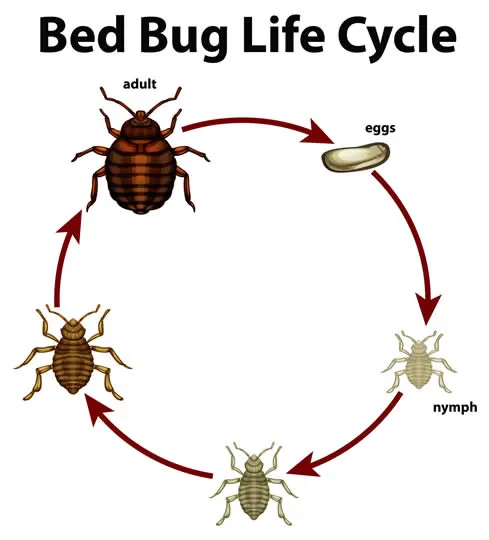
Bed bug bites are small, itchy, red bumps that result from the feeding of bed bugs, which are tiny insects that feed on human blood. Bed bug bites usually occur on exposed skin during sleep and may appear in clusters or lines. Bed bug bites are not dangerous, but they can cause allergic reactions or skin infections in some people.

bed

What are the signs and symptoms of Bed Bug Bites?

The signs and symptoms of bed bug bites are:

  • Red and swollen welts, often with a dark spot in the center, that may look like hives or welts
  • Itchy and burning sensations on the skin
  • Bites arranged in lines or clusters, usually on exposed skin during sleep
  • Blood stains on the bedding from scratching
  • Allergic reactions or skin infections in some people

What treatments are available at the dermatologist for Bed Bug Bites

The treatments available at the dermatologist for bed bug bites are:

  • Steroid ointments and antihistamines. These are the most common bedbug bite treatments. They relieve itchiness and swelling. Scratching bedbug bites can lead to skin infections.
  • Injection of an antihistamine, corticosteroid, or epinephrine. Some people may require an injection of one of these medications for a severe allergic reaction.
  • Professional extermination or heat treatment. It can be difficult to get rid of a bedbug infestation because they hide well and can live months without eating. You may need to hire a professional exterminator, who will likely use a combination of pesticides and nonchemical treatments. You may also treat your home for bedbugs by vacuuming, laundering, or placing items in an enclosed vehicle.
bed-bug-life-cycle

Bed Bug Bites vs Mosquito Bites

Bed bug bites and mosquito bites are both red, itchy, and may look similar. However, there are some differences between them that can help you identify what bit you. Some of the differences are:

  • Appearance: Bed bug bites usually appear in clusters or lines of three to five bites, often on exposed skin during sleep. Mosquito bites are usually isolated and random, and can occur on any part of the body that clothing does not cover.
  • Reaction time: Bed bug bites may not cause any skin reaction for hours or days after the bite, while mosquito bites are usually instantly itchy and visible.
  • Infection: Mosquito bites can transmit serious diseases such as malaria, dengue, Zika, and West Nile virus, while bed bug bites are not known to spread any diseases.

FAQ About Bed Bug Bites

Where are bed bugs found?

Bed bugs are found across the globe from North and South America, to Africa, Asia and Europe. Bed bugs have been found in five-star hotels and resorts and their presence is not determined by the cleanliness of the living conditions where they are found. Bed bug infestations usually occur around or near the areas where people sleep.

Do bed bugs spread disease?

Bed bugs are not known to spread disease. Bed bugs can be an annoyance because their presence may cause itching and loss of sleep.

How do I know if I’ve been bitten by a bed bug?

 It is hard to tell if you’ve been bitten by a bed bug unless you find bed bugs or signs of infestation. When bed bugs bite, they inject an anesthetic and an anticoagulant that prevents a person from realizing they are being bitten.

Is there a dermatologist near me in Austin that offers treatment for Bed Bug Bites?

Yes. At our Austin dermatology office we offer treatment for Bed Bug Bites to patients from Austin and the surrounding area. Contact our office today to schedule an appointment.